MT2 Dosage: Amounts Used in Melanotan 2 Studies
What amounts of Melanotan 2 (MT-II) did researchers use? This page lists the MT2 dosage figures printed in the study papers, and nothing more. People got a narrow range. Animals got other amounts, given in other ways. None of these numbers is advice for you, and MT-II has no approval for use in people like you.
What amounts the studies used
People got only a narrow range of amounts. The first human study was Dorr's, in 1996. It began at 0.01 mg/kg under the skin [1]. When you see mg/kg, read it as milligrams for every kilogram of a person's weight, so that first amount came to about 0.7 mg for a person weighing 70 kg. The amount then rose to 0.025 mg/kg, across five shots in ten days. Two of the three men had darker skin. They also had nausea, sleepiness, and erections that lasted 1–5 hours.
In 1998, Wessells used 0.025 mg/kg under the skin [2]. His ten volunteers had erection trouble that their doctors thought began in the mind. The team picked that amount because it had caused erections in Dorr's tanning study. In 2000, a larger trial gave it to 20 men [3]. Severe nausea followed 12.9% of the shots at 0.025 mg/kg. You can take that to mean the amount sat close to the most these men could stand.
Animal studies changed both the amount and the way the drug went in. The amounts and methods used in studies include these:
- 0.01–0.025 mg/kg under the skin. The human range, from Dorr 1996 and Wessells 1998/2000 [1][2][3].
- 20 micrograms for every kilogram, under the skin every 48 hours. Rats, nerve repair, Ter Laak 2003 [13].
- 225 ng in all, put into the brain. A nanogram, written ng, is a billionth of a gram. Mice, insulin, Heijboer 2005 [12].
- 0.1 to 1 nanomole into both sides of one brain area. A nanomole is a lab measure for an amount far too small to see. Mice, hunger, Eliason 2022 [6].
You can't turn these into a set number of milliliters. The volume depends on how much MT-II was mixed into the liquid. Dorr and Wessells measured by body weight, not by volume. Both 0.01 and 0.025 mg/kg caused effects, and in the human trials 0.025 mg/kg was close to the most the men could bear.
How long Melanotan 2 took to darken skin
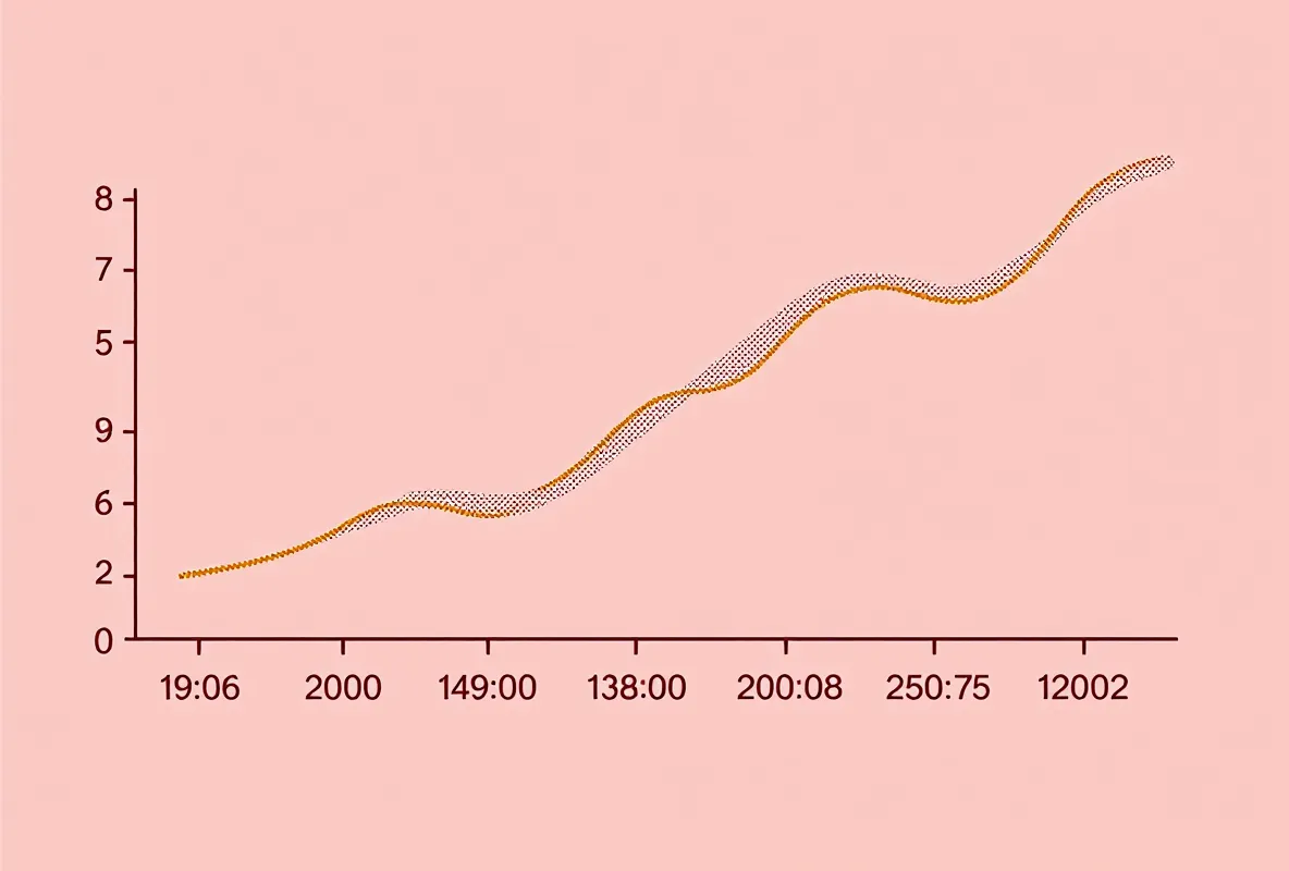
How long did it take? In the 1996 Dorr paper, five shots under the skin over ten days darkened the skin of two of the three men within 2–3 weeks [1]. A light meter measured the change, so you don't have to trust anyone's eyes. Your doctor can grade your skin by how it reacts to sun, on what's called the Fitzpatrick scale, and men in the lighter grades changed more. Color was already there at the first check after the shots. Three men were too few to show you when the color peaked.
What happened first after a study shot
What came first? In Dorr's 1996 trial, some effects began within 30–60 minutes [1]: nausea, flushing and erections. A shot under your skin can reach your blood and your brain inside an hour. Skin color took longer, and the light meter picked it up at 2–3 weeks. The drug's half-life in blood was about 1 hour. Half-life means the time it takes for half of it to leave your blood. The tan outlasted the drug because the skin cells had already made their dark pigment.
What the Melanotan 2 half-life tells you
How long does MT-II stay in your blood? About 1 hour is the best estimate. In 1994, Ugwu gave rats 0.3 milligrams for each kilogram of their weight, straight into a vein [16]. Two different lab tests of the rats' blood gave closely matching results. Too few late blood samples were taken, though, so you can't treat that figure as exact. Taken by mouth, only 4.6% reached the rats' blood, because the stomach and gut broke the rest apart.
In rodent blood, the natural tanning hormone lasts about 10–15 minutes. The ring shape and one swapped building block make MT-II last about 4–6 times longer. In studies of people given MT-I (afamelanotide) under the skin, its later half-life was 0.8–1.7 hours.
A tan can outlast the drug. Your skin cells make eumelanin, the dark pigment, and hand it on to the cells around them. Those cells stay until your skin sheds them. That's why the color in the Dorr study lasted past the last shot [1].
Time in your blood and time in your skin aren't the same thing. So how long MT-II stays in the blood tells you only part of when side effects may show up, since some effects may last after a dose has left the blood.
What researchers mixed before giving MT-II
What did the studies start with? A freeze-dried powder, which staff mixed with sterile water or sterile salt water. The papers say the dry powder keeps for a short time at room temperature and needs 4°C or colder, about the inside of your fridge, to keep longer. That comes from general rules for storing peptides, not from a test of MT-II itself. Dorr's 1996 paper describes its own mixing steps, and you won't find them turned into directions here. By mouth it barely gets in: only 4.6% got into the rats' blood by mouth [16]. That's why the human trials used shots beneath the skin.
What changed when the drug went in other ways
Animal studies tried four ways in: a shot under the skin, a shot into a vein, a dose straight into the brain, or drops up the nose. The human trials, Dorr 1996 and Wessells 1998/2000, used only shots under the skin [1][2][3]. You won't find any other way in tested for its effects in people.
In 2017, Paiva compared a vein shot with nose drops in rats [15]. The vein shot stirred up the brain cells that put out oxytocin, a bonding hormone. The nose drops didn't make those cells show the same signs of activity. The nose may not have carried enough MT-II into the rat brain.
In 2005 and 2003, Heijboer and Raposinho put MT-II straight into mouse or rat brains [12][14]. That let them tell brain effects apart from effects in the rest of the body.
You'd only see that route in careful animal work.
What makes the darker color last
Why does the color stay? In the 1996 Dorr study, darker skin stayed after the shots ended [1]. No human study has followed the fade on a set schedule. Animal work suggests the pigment builds up in the top layer of your skin and fades over weeks to months as old skin cells flake away. The skin switch tells cells to make pigment, and once the pigment is made, it doesn't vanish when MT-II leaves your blood. That's how a tan outlasts a drug with a blood half-life of ~1 hour.